An eye is like a little digital camera that relays what it sees to your brain, where the images are processed. In fact, many of the parts of your eye are imitated by a camera. Light passes through the pupil (aperture), and is focused by the lens. The amount of light let in is controlled by the iris (diaphragm). The focused image shines on the retina (charge-coupled device) and is converted to electrical signals which are sent to the brain (storage medium).
Let's look at all of the parts of the eye in greater detail.
Cornea
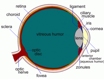
The cornea is the clear surface of the front of the eye. It helps direct light through the lens, so it contains no blood vessels. It is kept clean and moist by tears.
Anterior Chamber
This is the space between the cornea and iris, and is filled with a liquid called the Aqueous Humor.
Aqueous Humor
This fluid provides oxygen and nutrients to the cornea and iris.
Lens
The lens focuses light onto the retina at the back of the eye. The ciliary muscles and tiny fibres called zonules change its shape to let it focus on near or far objects.
Vitreous Humor
The vitreous humor is a gel that fills the rest of the eye.
Iris
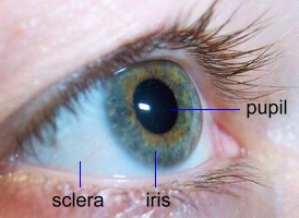
This is the coloured part of the eye; it's actually a ring of muscles that can contract or expand, making the pupil smaller or larger to let in less or more light.
Pupil
The pupil is the hole in the iris that light passes through.
|
See a real eye being dissected
|
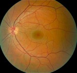 Retina
The retina lines the inside of the eyeball and is covered with light-sensitive nerves. It converts light into electrical signals and sends them to the brain through the optic nerve.
The sides of the retina are
responsible for our peripheral vision. The centre area, called the macula, is the most sensitive part of the retina; it registers light from objects we look directly at; it also is responsible for colour vision.
Retinal detachment occurs when the retina separates from the wall of the eye, and can no longer function. A detached retina can be put back where it belongs surgically, often by using a laser to 'weld' it back into place.
Fovea
The fovea is a tiny region in the macula that is the most sensitive to light. Because it is slightly off-centre, this means that your best vision is slightly to one side, rather than straight ahead. In full daylight you'll never notice the difference, but you can verify this by looking at stars late at night; stars that you look directly at are not quite as bright as when you look just to the left of them.
Optic Disk
The optic disk is the spot on the retina where the optic nerve exits the eye. There
are no sensory nerves here, so this spot inside the eye is a 'blind spot'. Each of your eyes has one, but each eye acts to cover for the blind spot
in the other eye, and the brain fills in the missing information ... so you don't notice the problem!
Optic Nerve
This is the central pathway where signals from all the sensory nerves inside the eye exit the eye and connect to the brain. Each optic nerve is made up of about 1.2 million nerve fibres.
 Conjunctiva
The conjunctiva is a thin, clear membrane covering the front of the eye; it produces mucous that helps lubricate the eye and protect it from infection.(Inflammation of this membrane is called conjunctivitis, or 'pink eye').
Choroid
The choroid is a layer of blood vessels between the retina and sclera that supplies blood to the retina.
Sclera
The sclera is the white, thick outer wall of the eye, which provides protection.
Visual Cortex
This is the part of the brain that processes the electrical signals from both eyes and converts them into 'pictures' that you can see.
Lacrimal Gland
This gland, (not shown on the diagram) more commonly known as the tear duct, releases protective fluids onto the surface of the eye to keep it moist.
Orbital Muscles
These six muscles (not shown on the diagram) surround the eyeball and control eye movement. Four of these can make the eye move up, down, left and right; the other two let the eye twist when we tilt our head.
Now let's have a look at how we actually see things ...
|
The Eye | How We See | Eye Problems | Dissecting an Eye
Resources
|