
In order to illustrate the parts of the eye a little more clearly, I decided to dissect an actual eyeball. We were able to obtain an elk eye; we chose this because of its large size, and also because it contains a tapetum lucidum layer that we wanted to examine.
Although larger than a human eye, the elk eye, except for the tapetum lucidum, is very similar, at least in terms of the main features.
|
 After slicing open the eyeball the vitreous humor began to leak out. It was a clear semi-liquid jelly that completely filled the interior of the eyeball.
|
|
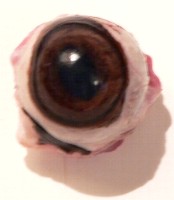
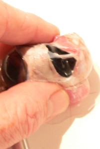
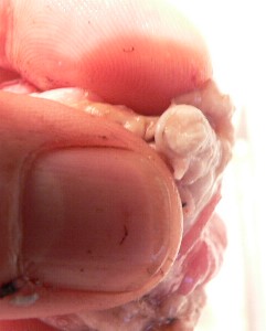
 The two photos above clearly show the cornea, a thin clear film that lines and protects the outside front of the eyeball. It was strong enough to prevent a finger from being poked through it. Notice also that the pupil is just a hole.
|
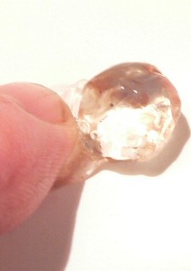
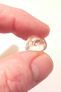
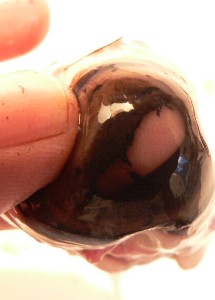
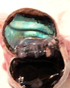
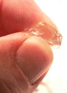 The lens was in the front of the eye right behind the pupil. Its surface was not a flattened ovoid shape, but was fairly bumpy and irregular. In the third photo you can see it in cross-section after it was cut in half. Surprisingly, its texture was much like wax, in that it could be squeezed out of shape if pressure was applied.
Exiting the back of the eye is the optic nerve; the second photo shows it in cross-section.
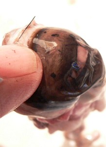
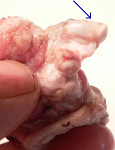
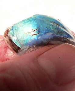
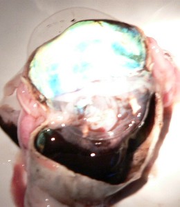
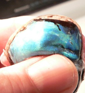 We examined the tapetum lucidum, a reflective layer at the back of the eye which acts like a mirror to increase the amount of light the photosensitive cells can collect. This allows many animals to see better under low-light conditions. The layer is a thin film stuck to the back of the eye, and is highly reflective. It is an iridescent blue colour in the elk eye. In the photos below, we stretched and sliced the layer so that you can see how thin it is. In the picture at the right below, we directed a flashlight at the layer in a dim room to make it clear that the layer really does relect light superbly.
The Eye | How We See | Eye Problems | Dissecting an Eye
Resources
|